


The anatomical structure of the venous system of the lower limbs is characterized by great diversity.Knowledge of the unique characteristics of the structure of the venous system plays a major role in the evaluation of instrumental examination data and in the selection of the correct treatment method.
The veins of the lower limbs are divided into superficial and deep.The superficial venous system of the lower limbs originates from the venous plexuses of the toes, forming the venous network of the back of the foot and the dorsal arch of the skin.This gives rise to the medial and lateral marginal veins, which enter the greater and lesser saphenous veins, respectively.The great saphenous vein is the longest vein in the body, it contains 5-10 pairs of valves, and its normal diameter is 3-5 mm.It originates in the lower third of the leg, in front of the medial epicondyle, and emerges in the subcutaneous tissue of the leg and thigh.In the groin area, the great saphenous vein flows into the femoral vein.Sometimes the great saphenous vein in the thigh and lower leg can be represented by two or even three trunks.The small saphenous vein begins in the lower third of the leg, along its lateral surface.In 25% of cases, it flows into the popliteal vein in the area of the popliteal fossa.In other cases, the small saphenous vein may rise above the popliteal fossa and drain into the femur, the great saphenous vein, or the deep femoral vein.
The deep veins of the posterior part of the leg begin with the dorsal metatarsal veins of the leg, which drain into the dorsal venous arch of the leg, from where blood flows into the anterior tibial veins.At the level of the upper third of the lower leg, the anterior and posterior tibial veins unite to form the popliteal vein, which is located laterally and slightly posterior to the artery of the same name.In the area of the popliteal fossa, the small saphenous vein and the veins of the knee joint flow into the popliteal vein.The deep vein of the thigh usually flows into the femoral vein 6-8 cm below the inguinal fold.Above the inguinal ligament, this vessel receives the epigastric vein, the deep vein surrounding the ilium, and passes into the external iliac vein, which joins the internal iliac vein at the sacroiliac joint.The paired common iliac vein begins after the confluence of the external and internal iliac veins.The right and left common iliac veins join to form the inferior vena cava.Large vessel without valves, 19-20 cm long and 0.2-0.4 cm in diameter.The inferior vena cava has parietal and visceral branches, through which blood flows from the lower limbs, lower trunk, abdominal organs and pelvis.
Perforating (communicating) veins connect the deep veins with the superficial ones.Most of them have valves located suprafascially, thanks to which blood flows from the superficial veins to the deep ones.There are direct and indirect perforating veins.The direct veins directly connect the deep and superficial venous networks, and the indirect veins flow indirectly, that is, they first flow into the muscular vein, which then flows into the deep vein.
The vast majority of perforating veins originate from tributaries rather than from the trunk of the great saphenous vein.In 90% of patients, the perforating veins of the medial surface of the lower third of the leg are incompetent.Incompetence of the perforating veins of Cockett, which connects the posterior branch of the great saphenous vein (Leonardo's vein) with the deep veins, is most often observed on the lower leg.In the middle and lower third of the thigh, there are usually 2-4 permanent perforating veins (Dodd, Gunter), which directly connect the trunk of the great saphenous vein with the femoral vein.In the case of varicose transformation of the small saphenous vein, incompetent communicating veins are most often observed in the middle and lower third of the leg and in the area of the lateral malleolus.
Clinical course of the disease
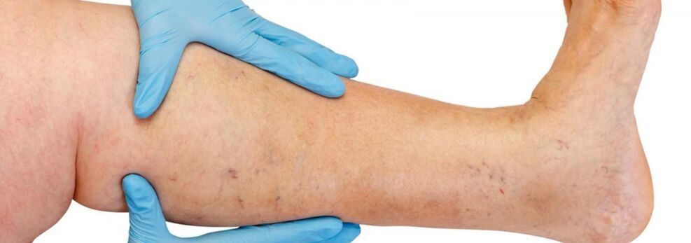
Varicose veins occur mostly in the saphenous system of the great vein, less often in the saphenous system of the small vein, and begin with the tributaries of the vein trunk on the legs.The natural course of the disease is quite favorable in the initial stage;for the first 10 years or more, patients should not be bothered by anything, apart from an aesthetic defect.After that, if the treatment is not carried out in time, the feeling of heaviness, tiredness of the legs and physical activity (long walk, standing) or swelling in the afternoon, especially after the hot season, begin to appear.Most patients complain of leg pain, but after detailed questioning it may be revealed that this is exactly the feeling of fullness, heaviness and fullness in the legs.Even with a short rest and the raised position of the limb, the severity of the sensations decreases.These symptoms characterize venous insufficiency at this stage of the disease.If we are talking about pain, other causes must be ruled out (arterial insufficiency of the lower limbs, acute venous thrombosis, joint pain, etc.).The later progression of the disease leads to the development of trophic disorders in addition to the increase in the number and size of dilated veins, often due to the development of incompetent perforating veins and the appearance of valvular insufficiency of the deep veins.
In case of insufficiency of perforating veins, trophic disorders are limited to any surface of the leg (lateral, medial, posterior).In the initial stage, trophic disorders manifest themselves in local hyperpigmentation of the skin, followed by thickening (induration) of the subcutaneous fat tissue until the development of cellulitis.This process ends with the formation of an ulcer-necrotic defect, which can reach a diameter of 10 cm or more and extends deep into the fascia.The typical location of venous trophic ulcers is the area of the medial malleolus, but the localization of ulcers on the leg can be different and multiple.In the stage of trophic disorders, severe itching and burning occur in the affected area;Some patients develop microbial eczema.The pain in the area of the ulcer is not pronounced, although in some cases it is intense.At this stage of the disease, the heaviness and swelling of the leg becomes permanent.
Diagnosis of varicose veins
Diagnosing the preclinical stage of varicose veins is especially difficult, since the legs of such patients may not have varicose veins.
In such patients, the diagnosis of varicose veins in the legs is wrongly rejected, although there are symptoms of varicose veins, there are indications that the patient's relatives suffer from this disease (hereditary tendency), as well as ultrasound data on the initial pathological changes in the venous system.
All of this can lead to missing the deadline for the optimal start of treatment, the development of irreversible changes in the vein wall, and the development of very serious and dangerous complications of varicose veins.Only if the disease is recognized in the early preclinical stage, it becomes possible to prevent pathological changes in the venous system of the legs through a minimal therapeutic effect on the varicose veins.
Avoiding various diagnostic errors and establishing the correct diagnosis is only possible after a thorough examination of the patient by an experienced specialist, a correct interpretation of all complaints, a detailed analysis of the medical history and maximum information obtained with the most modern tools (instrumental diagnostic methods) about the condition of the venous system of the legs.
A duplex scan is sometimes performed to determine the exact location of the perforating veins, color-coded to identify venous reflux.In case of valve insufficiency, their valves stop closing completely during the Valsava maneuver or compression test.Valvular insufficiency leads to the appearance of venous reflux, high through the incompetent saphenofemoral junction and low through the incompetent perforating veins of the leg.This method can be used to record the reverse flow of blood through the prolapsed leaflets of an incompetent valve.Therefore, the diagnosis is multi-stage or multi-level.In a normal situation, the diagnosis is made after ultrasound diagnostics and examination by a phlebologist.However, in particularly difficult cases, the examination must be carried out in stages.
- First, a phlebologist surgeon performs a thorough examination and questioning;
- if necessary, the patient is sent for additional instrumental research methods (duplex angioscanning, phleboscintigraphy, lymphoscintigraphy);
- patients with accompanying diseases (osteochondrosis, varicose eczema, lymphovenous insufficiency) are offered counseling with leading specialists about these diseases) or additional research methods;
- every patient requiring surgery is first consulted by the attending surgeon and, if necessary, by an anesthesiologist.
Treatment
Conservative treatment is mainly recommended for patients for whom surgical treatment is contraindicated: due to their general condition, in the case of mild dilatation of the veins causing only aesthetic discomfort, or in the case of rejection of surgical intervention.The aim of conservative treatment is to prevent further development of the disease.In these cases, patients should be advised to bandage the affected area with an elastic bandage or wear elastic stockings, regularly place their legs in a horizontal position, and perform special exercises on the feet and lower legs (flexion and extension in the ankle and knee joints) to activate the musculo-venous pump.Elastic compression accelerates and increases blood flow in the deep veins of the thigh, reduces the amount of blood in the saphenous veins, prevents the development of edema, improves microcirculation and helps normalize metabolic processes in the tissues.Bandaging should be started in the morning before getting out of bed.The bandage is applied by gently stretching it from the toes to the thigh, with the obligatory grip of the heel and ankle joint.Each subsequent round of knitting should overlap the previous one by half.It is recommended to use certified medical hosiery with an individual selection of the degree of compression (from 1 to 4).Patients should wear comfortable, hard-soled and low-heeled shoes, avoid prolonged standing, heavy physical work, and work in hot and humid places.If, due to the nature of the work activity, the patient has to sit for a longer period of time, the legs should be placed in an elevated position by placing a special stand of the appropriate height under the legs.It is advisable to walk a little every 1-1.5 hours or stand on tiptoes 10-15 times.The resulting contractions of the calf muscles improve blood circulation and increase venous outflow.While sleeping, the legs should be placed in an elevated position.
Patients are advised to limit water and salt intake, normalize their body weight, and regularly take diuretics and drugs that improve venous tone.According to the indications, drugs are prescribed that improve tissue microcirculation.The use of non-steroidal anti-inflammatory drugs is recommended for treatment.
Physiotherapy plays an important role in the prevention of varicose veins.For uncomplicated forms, water procedures are useful, especially swimming, warm (no higher than 35°) foot baths with 5-10% common salt solution.
Compression sclerotherapy
The indications for injection therapy (sclerotherapy) for varicose veins are still debated.The method consists of injecting a sclerosing agent into the dilated vein, its further compression, desolation and sclerosis.Modern drugs used for such purposes are quite safe, that is, they do not cause necrosis of the skin or subcutaneous tissue when administered extravasally.Some specialists use sclerotherapy for almost all forms of varicose veins, while others completely reject the method.The truth probably lies somewhere in the middle, and for young women in the initial stages of the disease, it makes sense to use the injection method of treatment.The only thing is that they should be warned about the possibility of relapse (greater than in the case of surgical intervention), the need to wear a fixed compression bandage for a long time (up to 3-6 weeks) and the possibility that several sessions may be required for complete sclerosis of the veins.
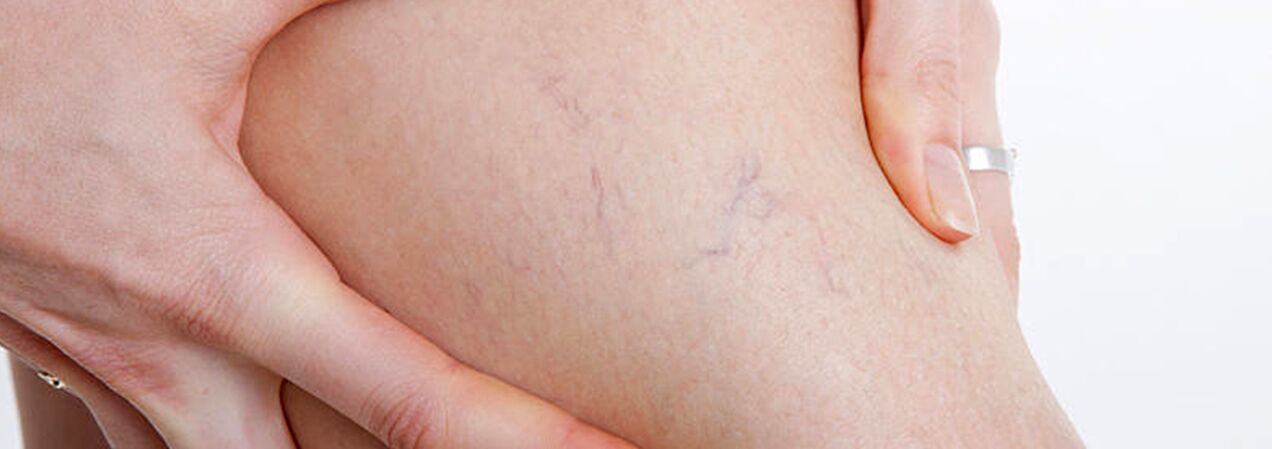
Patients suffering from telangiectasias ("spider veins") and reticular dilatation of the small saphenous veins should be included in the group of varicose veins, since the causes of the development of these diseases are the same.In this case, in addition to sclerotherapypercutaneous laser coagulation, but only after ruling out damage to the deep and perforating veins.
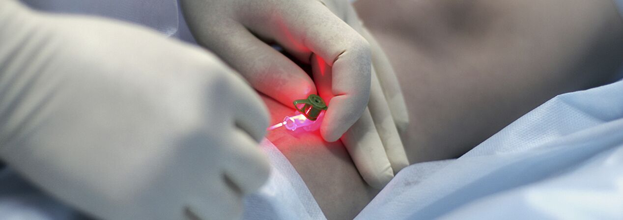
Percutaneous laser coagulation (PLC)
This is a method based on the principle of selective photocoagulation (photothermolysis) and the absorption of laser energy by various substances in the body.The specialty of the method is the non-contact nature of this technology.The focusing head concentrates the energy into the blood vessel in the skin.Hemoglobin in blood vessels selectively absorbs laser beams of a certain wavelength.As a result of the laser, the endothelium in the vessel lumen is destroyed, which leads to adhesion of the vessel walls.
The effectiveness of PLK directly depends on the penetration depth of the laser radiation: the deeper the vessel, the longer the wavelength must be, so the indications of PLK are quite limited.Microsclerotherapy is most effective for vessels with a diameter greater than 1.0-1.5 mm.Taking into account the extensive and branching distribution of spider veins on the legs and the varying diameter of the vessels, a combined treatment method is currently actively used: in the first stage, sclerotherapy is performed for veins with a diameter of more than 0.5 mm, and then the remaining "stars" with a smaller diameter are removed with a laser.
The procedure is practically painless and safe (no skin cooling or anesthesia is used), since the light of the device belongs to the visible part of the spectrum, and the wavelength of the light is designed so that the water in the tissues does not boil and the patient does not get burns.In case of high sensitivity to pain, prior application of local anesthetic cream is recommended.Erythema and swelling disappear within 1-2 days.After the course, for about two weeks, some patients may experience darkening or lightening of the treated skin area, which then disappears.For fair-skinned people, the changes are almost imperceptible, but for dark-skinned or heavily tanned patients, the risk of such temporary pigmentation is quite high.
The number of procedures depends on the complexity of the case - the veins are of different depths, the lesions may be smaller or occupy a rather large skin area, but usually no more than four laser treatments (5-10 minutes each) are required.The maximum result is achieved in such a short time by the unique "square" shape of the device's light pulse;compared to other devices, it increases its effectiveness, reducing the possibility of side effects after the intervention.
Surgical treatment
Surgery is the only radical treatment method for patients with varicose veins of the lower extremities.The aim of the operation is to eliminate the pathogenetic mechanisms (veno-venous reflux).This is accomplished by removing the main trunks of the great and small saphena and ligating the incompetent communicating veins.
The surgical treatment of varicose veins goes back a hundred years.Previously and many surgeons still used large incisions along the varicosities and used general or spinal anesthesia.The marks after such a "mini-phlebectomy" will remind you of the operation for a lifetime.The first operations on veins (according to Schade, according to Madelung) were so traumatic that the damage they caused exceeded the damage caused by varicose veins.
In 1908, the American surgeon Babcock developed a method of extracting subcutaneous veins using a rigid metal probe with an olive.In an improved form, the surgical method for removing varicose veins is still used in many public hospitals.Varicose veins are removed through a separate incision, as suggested by surgeon Narat.Thus, the classic phlebectomy is called the Babcock-Narat method.Phlebectomy according to Babcock-Narat has disadvantages - large postoperative scars and impaired skin sensitivity.The ability to work is reduced for 2-4 weeks, which makes it difficult for patients to agree to surgical treatment of varicose veins.
Phlebologists have developed a unique technology to treat varicose veins in one day.They operate with the help of complex casescombined technology.The main large varicose veins are removed by inversion stripping, which means a minimal intervention through mini-incisions of the skin (2-7 mm), which leave virtually no scars.The use of the minimally invasive technique involves minimal tissue trauma.The result of this operation is the elimination of varicose veins, with excellent aesthetic results.Combined surgical treatment is performed under full intravenous or spinal anesthesia with a maximum of 1 day of hospitalization.
Surgical treatment includes:
- Crossectomy - crossing the place where the trunk of the great saphenous vein flows into the deep venous system;
- Stripping is the removal of a fragment of a varicose vein.Only the varicose veins are removed, and not the whole (as in the classic version).
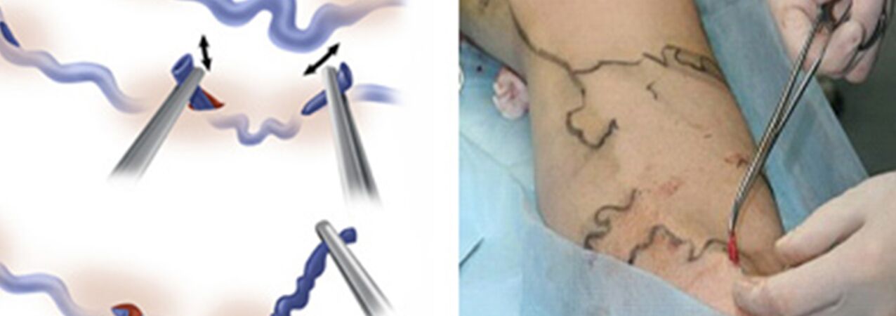
Actuallyminiphlebectomyreplaced the Narat technique for removal of varicose tributaries of major veins.Previously, 1-2-5-6 cm skin incisions were made along the varices, through which the veins were isolated and removed.The desire to improve the cosmetic result of the intervention and to be able to remove the veins not with a traditional incision, but with a mini-incision (puncture), forced doctors to develop devices that can do almost the same thing through a minimal skin defect.This is how phlebectomy "hooks" of different sizes and configurations, as well as special spatulas, appeared.And instead of the usual scalpel, scalpels with a very narrow blade or needles with a fairly large diameter began to be used to pierce the skin (for example, a needle for analyzing venous blood with a diameter of 18G).Ideally, the trace of a puncture with such a needle is practically invisible after a while.
Some forms of varicose veins are treated on an outpatient basis under local anesthesia.Minimal trauma during miniphlebectomy, as well as the low risk of the intervention, make it possible to perform this operation in a day hospital.After the operation, the patient can be sent home independently after minimal observation in the clinic.In the postoperative period, maintaining an active lifestyle, encouraging active walking.Temporary incapacity for work usually lasts a maximum of 7 days, after which you can work.
When is microphlebectomy used?
- If the diameter of the varicose trunks of the great or small saphenous vein exceeds 10 mm;
- After suffering from thrombophlebitis of the main subcutaneous trunks;
- After trunk recanalization after other types of treatments (EVLT, sclerotherapy);
- Removal of very large individual varicose veins.
It can be an independent operation or a component of the combined treatment of varicose veins, combined with laser treatment of veins and sclerotherapy.The tactics of use are determined individually, always taking into account the results of ultrasound duplex scanning of the patient's venous system.Microphlebotomy is used to remove veins in various places that have changed for various reasons, including on the face.Professor Varadi from Frankfurt developed his convenient instruments and formulated the basic postulates of modern microphlebectomy.The Varadi phlebectomy method provides excellent cosmetic results without pain or hospitalization.This is very laborious, almost jewelry work.
After vein surgery
The postoperative period after the usual "classic" phlebectomy is quite painful.Sometimes large hematomas cause concern and swelling occurs.Wound healing depends on the phlebologist's surgical technique;sometimes there is leakage of lymph and long-term formation of noticeable scars;often, after a major phlebectomy, a loss of sensation remains in the heel area.
In contrast, after miniphlebectomy, the wounds do not require suturing, since they are only punctures, there is no pain, and in practice no damage to the cutaneous nerve has been observed.However, such results of phlebectomy are achieved only by highly experienced phlebologists.